
Fig. 4
- ID
- ZDB-FIG-170125-13
- Publication
- Beretta et al., 2017 - Early Commissural Diencephalic Neurons Control Habenular Axon Extension and Targeting
- Other Figures
- All Figure Page
- Back to All Figure Page
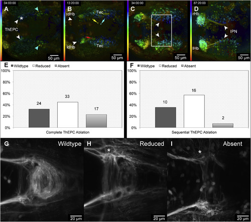
Complete and Incomplete Unilateral ThEPC Cell Ablations Cause Differently Severe Bilateral IPN Innervation Defects (A–D) Dorsal view, anterior to the left, color-coded MIP of four developmental stages acquired by in vivo 2-PM after sequential unilateral ThEPC cell ablation at 32 hpf in an Et(−1.0otpa:mmGFP)hd1 embryo. The LUT shows the z color-coded table according to the depth of each stack. Original stacks were cropped and gamma was adjusted to a value of 0.60 for display purposes. (A) For orientation, white arrowheads mark the position of ThEPC neurons; blue arrowheads mark the second posterior bilateral cluster of projecting neurons. An asterisk marks the ablated side. (B) White arrowheads mark the left and right dHb. The blue dot and arrow mark the tip of a commissural tectal axon; red and yellow dots and arrows mark ipsilateral and commissural ThEPC axons, respectively. (C and D) White arrowheads and box indicate the area in which dHb axon elongation stalls upon complete ThEPC cell ablation at (C) 66 hpf and (D) 119 hpf. (E and F) Statistics of IPN innervation phenotypes observed after (E) 74 independent unilateral complete and (F) 28 sequential ThEPC cell ablations. Numbers above the columns correspond to the number of embryos showing the respective phenotype. (G–I) Dorsal views with anterior to the left showing normal IPN innervation (G) and examples of the indicated IPN innervation phenotypes after sequential unilateral ThEPC neuron ablations (H and I). Asterisks in (H) and (I) mark the site of ablation. d, dorsal; Hb, habenula; IPN, interpeduncular nucleus; l, left; r, right; Tec, optic tectum; ThEPC, thalamic-epithalamic early projecting cluster. See also Figures S3 and S4 and Movie S3. |