
Fig. 6
- ID
- ZDB-FIG-170103-32
- Publication
- Montalbano et al., 2016 - Retinoic acid catabolizing enzyme CYP26C1 is a genetic modifier in SHOX deficiency
- Other Figures
- All Figure Page
- Back to All Figure Page
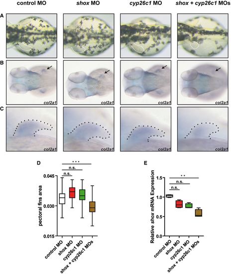
Co‐injection of titrated subphenotypic doses of anti‐shox and anti‐cyp26c1 morpholinos impairs limb development Wild‐type embryos injected with control MO, subphenotypic doses of shox MO, cyp26c1 MO, or a combination of shox + cyp26c1 MOs. A. Dorsal views of the embryos at 55 hours post‐fertilization (hpf). Dotted line, pectoral fins. shox + cyp26c1 double morphants show smaller fins compared to control and single MOs (n = 30 embryos). B, C Dorsal view and magnification on the pectoral fins of col2a1 expression at 55 hpf. Arrow and dotted line indicate the pectoral fin. D. Pectoral fin area was measured by ImageJ (n = 30 embryos). Data are shown as means ± SD. ***P = 0.0001 (one‐way ANOVA with Bonferroni's multiple comparison test). E. Relative expression of shox mRNA normalized to reference genes eef1a and b‐actin in zebrafish embryos injected with control MO, shox MO, cyp26c1 MO, or shox + cyp26c1 MOs (n = 4). RNA was extracted from 10–15 injected embryos at 55 hpf. Data are shown as means ± SD. **P = 0.0048 (Kruskal–Wallis with Dunn's multiple comparison test). |
| Gene: | |
|---|---|
| Fish: | |
| Knockdown Reagents: | |
| Anatomical Term: | |
| Stage: | Day 5 |
| Fish: | |
|---|---|
| Knockdown Reagents: | |
| Observed In: | |
| Stage Range: | Long-pec to Day 5 |