
Fig. 1
- ID
- ZDB-FIG-161229-5
- Publication
- James et al., 2016 - The Hyaloid Vasculature Facilitates Basement Membrane Breakdown During Choroid Fissure Closure in the Zebrafish Eye
- Other Figures
- All Figure Page
- Back to All Figure Page
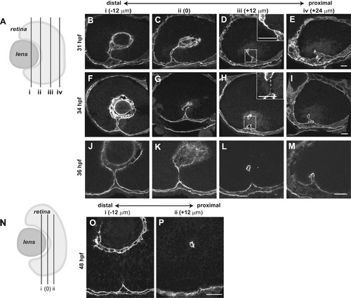
Temporal and spatial dynamics of basement membrane breakdown during choroid fissure closure in zebrafish. (A) Schematic depicting the approximate level of sections in B-M along the proximal-distal axis of the CF. The vitreous cavity was defined as central, and sections were taken at 12 µm intervals proximally and distally from this point. (B-M) Sagittal sections along the proximal-distal axis of the retina, immunostained for Lam-111 expression. (B-E) 31 hpf, (F-I) 34 hpf, (J-M) 36 hpf. Insets in D, H show high magnification views of the regions in the dashed boxes. (N) Schematic depicting the plane of section for 48 hpf embryos in O,P. (O,P) Representative sagittal sections along the proximal-distal axis of the retina immunostained for Lam-111. Scale bars=20 µm. |
Reprinted from Developmental Biology, 419(2), James, A., Lee, C., Williams, A.M., Angileri, K., Lathrop, K.L., Gross, J.M., The Hyaloid Vasculature Facilitates Basement Membrane Breakdown During Choroid Fissure Closure in the Zebrafish Eye, 262-272, Copyright (2016) with permission from Elsevier. Full text @ Dev. Biol.