
Fig. 7
- ID
- ZDB-FIG-161229-36
- Publication
- Galant et al., 2016 - Embryonic origin and lineage hierarchies of the neural progenitor subtypes building the zebrafish adult midbrain
- Other Figures
- All Figure Page
- Back to All Figure Page
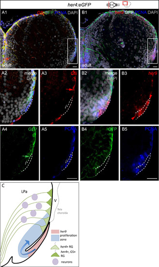
The sequence of progenitor subtypes at the lateral pallial edge is compatible with a reiteration of the TeO sequence. Compared expression of Her4-GFP, GS and PCNA (A) and her9, Her4-GFP and PCNA (B) in triple labelled cross-sections of the adult pallium, counterstained with DAPI. her9 expression is revealed by fluorescent in situ hybridization, and all other markers by immunocytochemistry. Confocal views. Panels 2–5 are high magnifications views of the boxed areas in panels 1. Panels 1 is the merged view, panels 3–5 are single channels. Red and green arrows in A3, A4 point to the lateral limit of the GS- and Her4-GFP-positive zones, respectively. The red arrowhead in B3 points to the medial limit of the her9-positive domain. Scale bars: 50 µm. C. Interpretative drawing showing the relative positions of the her9, PCNA, her4 and GS-positive zones. This organization is reminiscent of the TeO neurogenic sequence. Progression from a lateral pool to RG was shown in Dirian et al., (2014). |
Reprinted from Developmental Biology, 420(1), Galant, S., Furlan, G., Coolen, M., Dirian, L., Foucher, I., Bally-Cuif, L., Embryonic origin and lineage hierarchies of the neural progenitor subtypes building the zebrafish adult midbrain, 120-135, Copyright (2016) with permission from Elsevier. Full text @ Dev. Biol.