
Fig. 6
- ID
- ZDB-FIG-161214-6
- Publication
- Hanashima et al., 2017 - Complete primary structure of the I-band region of connectin at which mechanical property is modulated in zebrafish heart and skeletal muscle
- Other Figures
- All Figure Page
- Back to All Figure Page
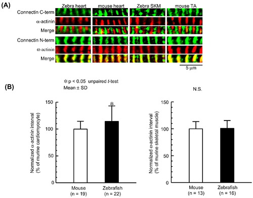
Comparison of the sarcomere lengths in heart and skeletal muscle between mouse and zebrafish. (A) Immunostaining of zebrafish heart and skeletal muscle using connectin N-terminus or C-terminus and α-actinin antibodies. Mouse heart and tibialis anterior muscle (TA) were also stained as controls. The localizations of N-terminus connectin at the Z-line and C-terminus connectin at the M-line are conserved in heart and skeletal muscle of zebrafish and mouse. Green, connectin. Red, α-actinin. SKM, skeletal muscle. (B) The sarcomere lengths of zebrafish heart and skeletal muscle, which are normalized to those of mouse heart and tibialis anterior muscle, respectively. The sarcomeres of zebrafish heart are longer than those of mouse heart, and those of zebrafish skeletal muscle are similar to those of mouse tibialis anterior muscle. Asterisk indicates p < 0.05 in t-test. |
| Antibodies: | |
|---|---|
| Fish: | |
| Anatomical Terms: | |
| Stage: | Adult |
Reprinted from Gene, 596, Hanashima, A., Hashimoto, K., Ujihara, Y., Honda, T., Yobimoto, T., Kodama, A., Mohri, S., Complete primary structure of the I-band region of connectin at which mechanical property is modulated in zebrafish heart and skeletal muscle, 19-26, Copyright (2017) with permission from Elsevier. Full text @ Gene