
Fig. S2
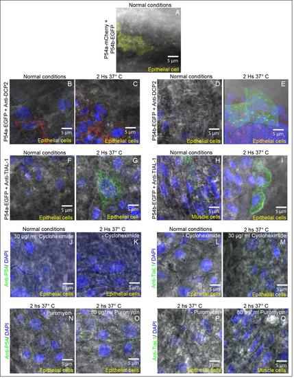
Differential interference contrast (DIC) illumination and cytoplasmic granules in epithelial and muscle cells. In all images we combined DIC illumination and fluorescence microscopy. Nuclei were labeled with DAPI (blue labeling) and cytoplasmic granules with different labels.(A) Cytoplasmic granules of P54a-mCherry and P54b-EGFP fusion reporters in an epithelial cell under normal conditions (same as Fig. 2K). (B and C) Cytoplasmic P54a-EGFP- and anti-Dcp2 labeled granules in an epithelial cell under normal conditions (B same as Fig. 2N) and in heat shock conditions (C same as Fig. 2Q). (D and E) P54b-EGFP- and anti-Dcp2 in cytoplasmic granules in an epithelial cell under normal conditions (D same as Fig. 2T) and in heat shock conditions (E same as Fig. 2W). (F and G) Epithelial cells with cytoplasmic granules containing P54a-EGFP- and labeled with anti-TIAL-1, in normal (F same as Fig. 3G) or heat shock conditions (G same as Fig. 3J). (H) Muscle cell and (I) Epithelial cells, both with cytoplasmic granules containing P54b-EGFP- and anti-TIAL-1, in normal (same as Fig. 3M) or heat shock conditions (same as Fig. 3P), respectively. (J and K) Anti-P54 labeling cytoplasmic granules, in epithelial cells, treated with or without cycloheximide and in normal temperature conditions (same as Fig. 4B) or in heat shock (same as Fig. 4C), respectively. (L and M) Cycloheximide treated and untreated epithelial cells, with anti-P54 labeling cytoplasmic granules, in normal conditions (same as Fig. 4E) or in heat shock (same as Fig. 4H), respectively. (N and O) Cytoplasmic granules labeled with anti-P54 antibody, in epithelial cells, treated with or without puromycin, both in heat shock conditions (same as Fig. 4K and L respectively). (P) Epithelial cell where cytoplasmic granules were labeled with anti-TIAL-1 antibody (same as Fig. 4O) and exposed to a heat shock. (Q) Muscular cell treated with puromycin and in heat shock conditions where cytoplasmic granules were labeled with anti-TIAL-1 antibody. All these images are the same as some Figures 2, 3 and 4 and are presented here with overlapping DIC illumination to observe the type of cell studied. |