
Fig. 5
- ID
- ZDB-FIG-161116-5
- Publication
- Rebman et al., 2016 - Cadherin-2 Is Required Cell Autonomously for Collective Migration of Facial Branchiomotor Neurons
- Other Figures
- All Figure Page
- Back to All Figure Page
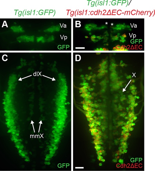
Expression of dominant-negative Cadherin-2 in trigeminal and vagus branchiomotor neurons leads to aberrant neuron positioning. (A) Dorsal view of live Tg(isl1:GFP) embryo at 48hpf shows positioning of anterior and posterior clusters of trigeminal neurons (Va,Vp) found in r2 and r3, respectively. Note the lateral positioning of the Va cluster of trigeminal motor neurons at 48 hpf. (B) Dorsal view of live Tg(isl1:GFP)/Tg(isl1:cdh2ΔEC-mCherry)vc25 embryo at 48 hpf shows that trigeminal neurons (Va; asterisk) remain in a medial location. Green is the GFP signal, whereas red is the Cdh2ΔEC-mCherry signal. (C) Dorsal view of live Tg(isl1:GFP) embryo at 48hpf shows correct positioning of vagus motor neurons in dorsolateral motor nucleus (dlX) and medial motor nucleus (mmX). (D) Dorsal view of live Tg(isl1:GFP)/Tg(isl1:cdh2ΔEC-mCherry)vc25 embryo shows that vagus neurons (X) do not migrate and coalesce into discrete dorsolateral nuclei. Scale Bars = 20 μm. |