
Fig. 6
- ID
- ZDB-FIG-161103-12
- Publication
- Sicca et al., 2016 - Gain-of-function defects of astrocytic Kir4.1 channels in children with autism spectrum disorders and epilepsy
- Other Figures
- All Figure Page
- Back to All Figure Page
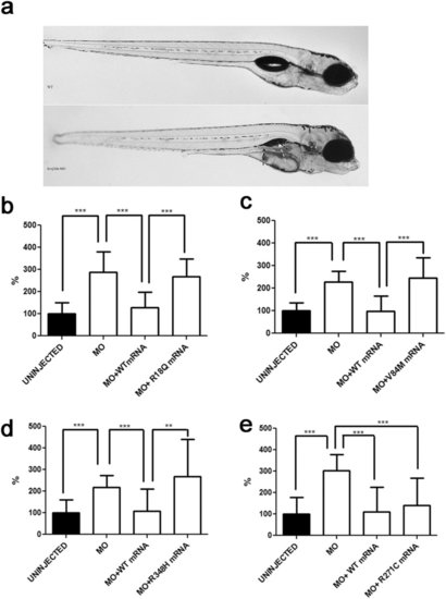
In vivo modelling of Kir4.1 mutations in zebrafish. (a) Transient kcnj10a knockdown zebrafish show macroscopic abnormalities in organ development compared to wild-type (WT). In morphant embryos, the pronephric duct is visible because it is dilated (black arrow); the swim bladder is not visible (white arrow). (b-e) Number of spontaneous tail flicks (registration time 30sec) seen in WT and mutant 30 hpf embryos. Values are expressed as percent of flicks counted in uninjected embryos. Embryos injected with MO (b-e), and with equimolar amount of MO and either R18Q (b), V84M (c), and R348H (d) mRNA show an increased rate of spontaneous contractions, compared to uninjected embryos (b-e), and to either MO+WT (b-e) and MO+R271C (e) mRNA injected embryos. **p < 0.01; ***p < 0.001. |
| Fish: | |
|---|---|
| Knockdown Reagent: | |
| Observed In: | |
| Stage: | Prim-15 |