
FIGURE
Fig. 7
Fig. 7
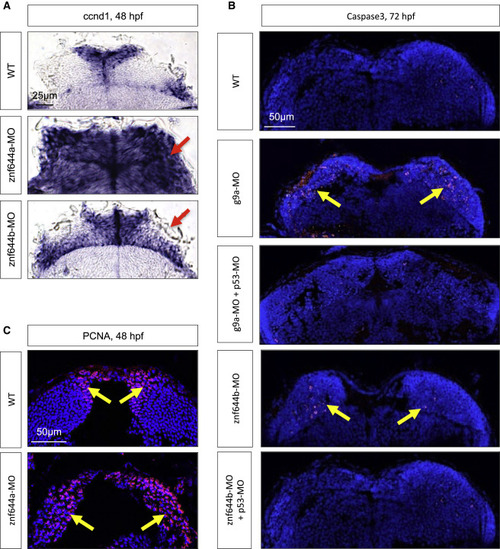
The Retinal Defects of g9a, znf644a, and znf644b Morphants Are Recapitulated in the Midbrain (A) WISH assays monitoring ccnd1 expression in WT, znf644a, or znf644b morphant midbrain cross-sections at 48 hpf. Arrows indicate mislocalized ccnd1 expression. (B) Immunostaining of cleaved Caspase3 in g9a or znf644b morphant midbrain cross-sections at 72 hpf and rescue by p53-MO (n = 3 in each group). Arrows denote the position of Caspase3+ cells. (C) Immunostaining of PCNA in WT or znf644a morphant midbrain cross-sections (n = 3 in each group). Arrows denote the position of PCNA+ cells. |
Expression Data
| Gene: | |
|---|---|
| Fish: | |
| Knockdown Reagents: | |
| Anatomical Term: | |
| Stage: | Long-pec |
Expression Detail
Antibody Labeling
Phenotype Data
| Fish: | |
|---|---|
| Knockdown Reagents: | |
| Observed In: | |
| Stage Range: | Long-pec to Protruding-mouth |
Phenotype Detail
Acknowledgments
This image is the copyrighted work of the attributed author or publisher, and
ZFIN has permission only to display this image to its users.
Additional permissions should be obtained from the applicable author or publisher of the image.
Full text @ Stem Cell Reports