
Fig. 3
- ID
- ZDB-FIG-160928-23
- Publication
- Jahr et al., 2016 - eduSPIM: Light Sheet Microscopy in the Museum
- Other Figures
- All Figure Page
- Back to All Figure Page
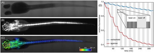
72 hpf zebrafish embryos expressing GFP in the vascular system. Prior to imaging, all zebrafish embryos were fixed and their fluorescence signal was recovered using booster-GFP. (A): Transmission image and (B): fluorescence image of vasculature of the same embryo. (C): Overlay of fluorescence and transmission data. Fluorescence data was 3D-rendered and colour coded for depth. Dataset of 75planes, colour bar, 500 m long. (D): Fluorescence signal of the sample embedded in vectashield (blue) and agarose (red). The sample was illuminated every 30s for 15min, followed by a 15min dark period (inset). Photobleaching dominated during bright periods. For mounting in vectashield, overall photobleaching was greatly reduced and even negligible during dark periods. |