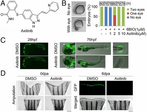
Axitinib inhibits Wnt/β-catenin signaling in zebrafish. (A) Chemical structure of axitinib. (B) Zebrafish embryos at 6 h post fertilization (hpf) were treated with 6BIO and axitinib at the indicated concentrations, and the eyeless phenotype was assessed 24 h later. n = total number of assessed embryos over four independent experiments. Embryos that died during treatment (n = 0, 4, 2, 0, 1, and 2 in the groups from left to right) were excluded from assessment. (C, Left) Representative images at 28 hpf of TCF-GFP transgenic zebrafish embryos treated with DMSO or axitinib (5 µM) for 2 d. n = 40 in each group; no embryos died. (Right) Representative images at 76 days post fertilization (dpf). The middle-hindbrain boundary (white arrows) and caudal fin mesenchyme are enlarged at the left and right, respectively. (D) Representative images of tailfin regeneration in TCF-GFP transgenic zebrafish (13 wk, n = 5 fish per group) treated with DMSO or axitinib (5 µM) for 6 d post amputation (dpa). Scale bars, 200 µm.
|
