
Fig. 4
- ID
- ZDB-FIG-160914-7
- Publication
- Taylor et al., 2016 - Zinc transporter ZIP10 forms a heteromer with ZIP6 which regulates embryonic development and cell migration
- Other Figures
- All Figure Page
- Back to All Figure Page
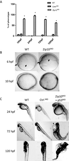
Phenotype of Zip10 knockdown zebrafish embryo Zebrafish embryos were co-injected with 2 ng of translation blocking morpholino-modified oligonucleotides against zip10 and p53 (to block off-target effects causing apoptosis; Zip10MO) or with 2 ng of a scrambled control morpholino (CtrlMO) and compared with uninjected wild-type (WT) embryos. (A) Percentage of embryos displaying morbidities (% phenotypes) at 10, 24, 72 and 120 hpf. (B) Epiboly cell migration in wild-type embryos and in embryos injected with a translation blocking morpholino against zip10 (Zip10MO) or with a scrambled control morpholino (CntrlMO). The extension of epipoly migration was delayed in Zip10MO embryos compared with time-matched controls (arrows) at 6 and 10 hpf with resulting delay in tail bud formation in morphants at 10 hpf compared with wild type. Morphant was yet to complete epiboly (85% complete) at 10 hpf when tail bud has already formed in wild type. (C) Common phenotypes of Zip10MO embryos at later stages of development (24-120 hpf). Note the Zip10MO embryo at 24 hpf with shorter anterior-posterior axis with abnormal head and eye formation (highlighted with white broken circle). Also note the oedema of the pericardial sac (black arrow-head) and the miss-formed heart (heart-string) feature (white arrow) in 72 hpf Zip10MO embryos. Severe oedema of pericardium and yolk sac was observed in many embryos at 120 hpf. Note also the curled or twisted tail at 72 and 120 hpf (white broken circles). The experiment was carried out five times with 20-50 embryos each time (N=5). Results were also replicated using a splicing blocking morpholino against zip10 (results not shown). |
| Fish: | |
|---|---|
| Knockdown Reagents: | |
| Observed In: | |
| Stage Range: | Shield to Day 5 |