
Fig. S2
- ID
- ZDB-FIG-160714-10
- Publication
- Scheid et al., 2016 - Essential light chain S195 phosphorylation is required for cardiac adaptation under physical stress
- Other Figures
- All Figure Page
- Back to All Figure Page
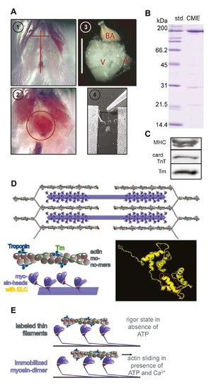
Preparation of actin and myosin for IVMA. (A) Horizontal skin section between the gills and a longitudinal section above the sternum (1) exposed the heart (2). After permeabilization and washing steps (3, scale bar 1mm), bulbus arteriosus (BA) and atrium (A) were removed, and only theventricle (V) was directly extracted in the IVMA flowchamber (4). (B) Protein contents of cardiac myosin extracts (CME) were analyzed via SDS-PAGE stained with Coomassie. Three main protein bands were present: one at around 200 kDa (myosin heavy chain, MHC), that was also detected by a specific antibody (AB) in WB (C), one at around 21 kDa (supposedly myosin light chain 1, MLC1) and around 14 kDa (supposedly MLC2). (C) Western blot analysis of the cardiac myosin extracts revealed the presence of the regulatory actin-connected proteins tropomyosin (Tm) and troponin T (TnT) in CME. (D) Before extraction of cardiac proteins, actin, myosin and regulatory proteins are arranged in interdigitating filaments within the sarcomere (upper part). The thin actin-containing filaments consist of actin monomers arranged in a double-helical filament (lower left panel). Filamentous Tm winds around the actin filament and is connected end-to-end via the troponin-complex. Myosin heads containing the myosin light chains protrude from the thick filament. The secondary protein structure of ELC is depicted as a ribbon model, encircled in red is the 11 amino acid part missing in the laz mutant (lower right panel; displayed via PyMOL, DeLano Scientific LLC, 2008; based on sequence from 9). (E) Schematic of IVMA: When myosin is extracted into IVMA flowchambers (A,4), the dissociated dimers are immobilized on a nitrocellulose-coated surface and will bind labeled thin filaments in a rigor-like state (upper panel). When ATP and Ca2+ are present, actomyosin cross-bridge cycling will lead to sliding of actin filaments (lower panel). |