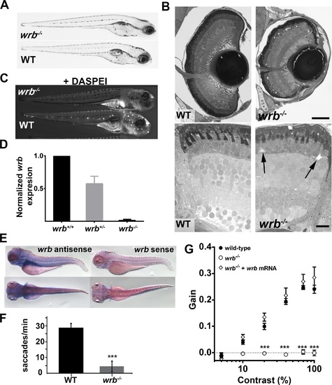
Visual system defects caused by retroviral insertion within the wrb gene. (A) Lateral views of 5 dpf wrb-/- and wild-type larvae. (B) Toluidine blue–stained 1-µm thick retinal sections from 5 dpf wild-type and wrb-/- larvae. Occasional acellular holes were observed in the ONL of wrb-/- mutants (arrows). (C) Live 5 dpf wrb-/- (top) and wild-type (bottom) larvae stained with the vital styryl dye DASPEI to label neuromasts. Two overlapping images, focused at the head or trunk, were stitched together for each panel. (D) Expression levels of wrb mRNA in heterozygous (wrb+/-) and mutant (wrb-/-) larvae at 5 dpf, compared with wild-type larvae, and normalized against beta-actin as measured by qRT-PCR. (E) Whole mount in situ hybridization at 5 dpf with antisense (left) and sense (right) wrb probes. (F) Saccade frequency measured from wild-type and wrb-/- larvae at 5 dpf. (G) Contrast response function measured from smooth pursuit eye movements. Gain versus log contrast for 5 dpf wild-type (black closed circles) wrb-/- (open circles) and wrb-/- rescued with wrb mRNA (open diamonds). Errors bars: SEM. Significance levels are as follows: ***P < 0.0001. Scale bars: 50 µm (B, top) 10 µm (B, bottom).
|
