
Fig. 3
- ID
- ZDB-FIG-160708-14
- Publication
- Koudelka et al., 2016 - Individual Neuronal Subtypes Exhibit Diversity in CNS Myelination Mediated by Synaptic Vesicle Release
- Other Figures
- All Figure Page
- Back to All Figure Page
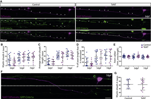
TeNT Expression in CoPA Neurons Does Not Impair Myelination along Individual Axons (A) Individual CoPA axons labeled with GFP-cntn1a and TdTomato (left) and with GFP-Cntn1a and TeNT-TdTomato (right) at 3 dpf. Scale bar, 10 µm. (B-E) Quantification of myelin sheath number per axon (B), average length of myelin sheath per axon per 425-µm imaging window (C), percentage of axon length (per 425-µm imaging window) that is myelinated (D), and axon caliber (E) at 3 dpf, 5 dpf, and 7 dpf in control and TeNT expressing CoPA neurons. (F) Individual CoPA neuron and axon labeled with GFP-Cntn1a and TeNT-TdTomato. Dashed line indicates dorsoventral cutoff for axonal region analyzed when assessing region of CoPA axons in ventral spinal cord. Scale bar, 15 µm. (G) Percentage of axon length that is myelinated in the ventral spinal cord of control and TeNT expressing CoPA neurons at 7 dpf. All error bars indicate ± SD. See also Figure S3. |