
FIGURE
Fig. 2
- ID
- ZDB-FIG-160630-55
- Publication
- Justice et al., 2016 - Evaluation of IRX Genes and Conserved Non-coding Elements in a Region on 5p13.3 Linked to Families with Familial Idiopathic Scoliosis and Kyphosis
- Other Figures
- All Figure Page
- Back to All Figure Page
Fig. 2
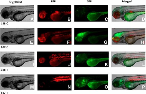
Patterns of GFP expression in F1 embryos at 72 hpf. Images of lateral views of representative embryos for each of the four constructs are shown in Brightfield (A, E, I, and M), RFP expression (B, F, J, and N), GFP expression (C, G, K, and O) and merged image of all three images (D, H, L, and P). In all images, embryos are oriented with their anterior to the left. |
Expression Data
| Genes: | |
|---|---|
| Fish: | |
| Anatomical Terms: | |
| Stage: | Protruding-mouth |
Expression Detail
Antibody Labeling
Phenotype Data
Phenotype Detail
Acknowledgments
This image is the copyrighted work of the attributed author or publisher, and
ZFIN has permission only to display this image to its users.
Additional permissions should be obtained from the applicable author or publisher of the image.
Full text @ G3 (Bethesda)