
Fig. 1
- ID
- ZDB-FIG-160630-40
- Publication
- Burger et al., 2016 - Maximizing mutagenesis with solubilized CRISPR-Cas9 ribonucleoprotein complexes
- Other Figures
- All Figure Page
- Back to All Figure Page
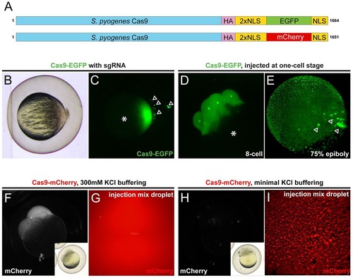
Microinjection of fluorescent Cas9 fusion proteins into zebrafish embryos. (A) Cas9 fusion proteins used in this study, showing the hemagglutinin tag (HA), two nuclear localization signals (NLS), EGFP or mCherry fluorophores, and a third C-terminal NLS. See Fig. S1 for protein sequences. (B,C) One-cell stage zebrafish embryo following microinjection of reconstituted Cas9-GFP-sgRNA complex. Brightfield (B) and EGFP fluorescence (C) reveal precipitated Cas9 complex outside the embryo (arrowheads, C), and strong EGFP fluorescence in the embryo cell without contribution to the yolk cell (asterisk, C). (D,E) Time-course of injected Cas9-EGFP and nuclear localization. Note the even distribution in the developing embryo without yolk fluorescence (asterisk, D) and minimal precipitate inclusions (arrowheads, E). (F-I) Buffering of the Cas9 protein-sgRNA mix with 300mM KCl maintains solubility in the injection mix (F,G) and prevents precipitation before injection (H,I). Insets (F,H) show corresponding brightfield images of fluorescence views. |