
Fig. 7
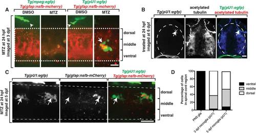
Ectopically located MEP glia do not elicit an immune response. A: Images of control and MTZ-treated Tg(mpeg:egfp);Tg(gfap:nsfB-mcherry) and Tg(pU1:egfp);Tg(gfap:nsfB-mcherry) larvae at 3 dpf. Arrowheads denote macrophages outside of the spinal cord. Arrow denotes microglia inside the spinal cord. Dashed lines denote the edge of the spinal cord. B: Transverse sections through Tg(pU1:egfp) embryos that were fixed at 5 dpf and stained with an antibody specific to acetylated tubulin. Arrow denotes location of microglia. C: Images of a Tg(pU1:egfp);Tg(gfap:nsfB-mcherry) embryo treated with MTZ at 24 hpf and imaged at 5 dpf. Arrows denote microglia. Note the lack of mcherry+ debris. D: Quantification of the location of microglia and ectopically located MEP glia within the spinal cord in MTZ-treated embryos at 3 and 5 dpf. Scale bars, 25 µm. |