
FIGURE
Fig. S1
Fig. S1
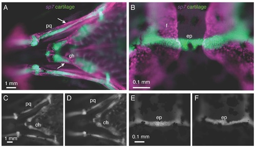
Cartilage does not express sp7 and is not affected in sp7 mutants. A, B) In a 3 wpf fish doubly transgenic for sp7:mCherry and col1a1:egfp, there is no overlap in expression of the two transgenes. A) Perichondral cells surrounding the palatoquadrate (pq) and ceratohyal (ch) cartilages (arrows) express the sp7 transgene. B) Similarly, the epiphyseal bar cartilage (ep) and the frontal bones (f) show no overlap in transgene expression. C-F) At 3 wpf, col1a1:egfp transgenics show no significant differences in cartilage formation or morphology between WT fish (C,E) and sp7 mutant (D,F). Scale bars are 1 mm. |
Expression Data
| Genes: | |
|---|---|
| Fish: | |
| Anatomical Terms: | |
| Stage: | Days 21-29 |
Expression Detail
Antibody Labeling
Phenotype Data
| Fish: | |
|---|---|
| Observed In: | |
| Stage: | Days 21-29 |
Phenotype Detail
Acknowledgments
This image is the copyrighted work of the attributed author or publisher, and
ZFIN has permission only to display this image to its users.
Additional permissions should be obtained from the applicable author or publisher of the image.
Reprinted from Developmental Biology, 413(2), Kague, E., Roy, P., Asselin, G., Hu, G., Stanley, A., Albertson, C., Simonet, J., Fisher, S., Osterix/sp7 limits cranial bone initiation sites and is required for formation of sutures, 160-72, Copyright (2016) with permission from Elsevier. Full text @ Dev. Biol.