
Fig. S5
- ID
- ZDB-FIG-160426-8
- Publication
- Berg et al., 2016 - Lysosomal Disorders Drive Susceptibility to Tuberculosis by Compromising Macrophage Migration
- Other Figures
- All Figure Page
- Back to All Figure Page
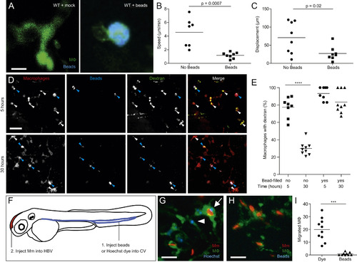
Lysosomal Accumulation of Inert Particles Compromises Endocytic Recycling and Migration to the Initial Site of Mycobacterial Infection, Related to Figure 6 (A) Confocal images of green fluorescent macrophages in larvae mock-injected or injected with 5x10⁵ blue fluorescent 1 µm polystyrene beads. Scale bar, 12 µm. (B and C) Speed (B) and displacement (C) of macrophages with and without beads. (D) Confocal images of red fluorescent macrophages in 3dpf larvae pre-loaded with blue fluorescent polystyrene beads as in (A), injected 12 hr later with green fluorescent dextran and imaged at 5 and 30 hr post-dextran injection. Blue and white arrowheads denote macrophages containing dextran, with and without blue beads, respectively. Scale bar, 50 µm. (E) Quantification of macrophages that retained dextran at 5 and 30 hr post injection. (F) Diagram showing the experimental outline in which 2 dpf larvae were injected with Hoechst dye or beads in the CV followed by infection in the HBV with 200 Mm. (G and H) Confocal images of larval HBV containing green-fluorescent macrophages following CV injections with Hoechst (G) or blue fluorescent beads (H). Arrow and arrowhead denote Hoechst-positive macrophages that have migrated from the CHT, with and without phagocytosed red fluorescent Mm, respectively. Scale bar, 10 µm. (I) Number of macrophages in the HBV after injection of dye or beads in the CV followed by Mm infection in the HBV. Statistical significance was assessed using Student’s t test (B, C, and I), and one-way ANOVA with Sidak’s post test (E). |
Reprinted from Cell, 165, Berg, R.D., Levitte, S., O'Sullivan, M.P., O'Leary, S.M., Cambier, C.J., Cameron, J., Takaki, K.K., Moens, C.B., Tobin, D.M., Keane, J., Ramakrishnan, L., Lysosomal Disorders Drive Susceptibility to Tuberculosis by Compromising Macrophage Migration, 139-152, Copyright (2016) with permission from Elsevier. Full text @ Cell