
Fig. 1
- ID
- ZDB-FIG-160425-1
- Publication
- Nishioka et al., 2016 - ATX-LPA1 axis contributes to proliferation of chondrocytes by regulating fibronectin assembly leading to proper cartilage formation
- Other Figures
- All Figure Page
- Back to All Figure Page
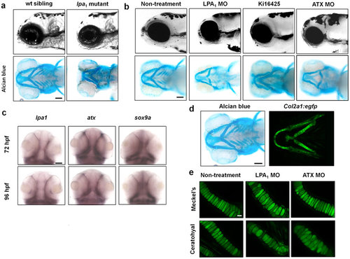
Loss of ATX-LPA1 signaling resulted in dyschondroplasia in zebrafish. (a,b) Loss of ATX- LPA1 signaling leads to deformation of the cephalic region in zebrafish embryos. Cephalic regions of wt and lpa1 mutant zebrafish embryos (a) and cephalic regions of zebrafish embryos treated with LPA1 antagonist, LPA1 or ATX morpholinos (b) at 96 hpf (side view). Cartilage tissues visualized by alcian blue and alizarin red staining are also shown (ventral view). Scale bar: 100 µm. (c) Both lpa1 and atx are co-expressed with sox9, a marker of chondrocytes, in zebrafish embryo both at 72 and 96 hpf as judged by in situ hybridization. Scale bar: 50 µm. (d,e) Loss of ATX-LPA1 signaling in zebrafish embryos leads to mislocalization of chondrocytes in cartilage tissues. (d) EGFP is expressed specifically in chondrocytes in col2:egfp transgenic zebrafish at 120 hpf. Scale bar: 100 µm. (e) In zebrafish embryos treated with LPA1 or ATX morpholinos, chondrocytes are unevenly distributed in Meckel’s and ceratohyal cartilages at 120 hpf. Scale bar: 100 µm. |
| Genes: | |
|---|---|
| Fish: | |
| Anatomical Term: | |
| Stage Range: | Protruding-mouth to Day 5 |
| Fish: | |
|---|---|
| Condition: | |
| Knockdown Reagents: | |
| Observed In: | |
| Stage Range: | Day 4 to Day 5 |