
Fig. 1
- ID
- ZDB-FIG-160323-11
- Publication
- Lin et al., 2015 - Ras-Related Nuclear Protein is required for late developmental stages of retinal cells in zebrafish eyes
- Other Figures
- All Figure Page
- Back to All Figure Page
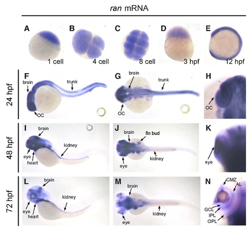
The spatiotemporal expression pattern of ran mRNA in developing zebrafish embryo. (A-E) From one-cell stage up to 12 hpf, ran mRNA was detected on the whole embryo. (A,D,E) Lateral view; (B,C) animal polar view. (F-H) At 24 hpf, while ran mRNA can still be detected in brain and optic cup, the level of ran mRNA started to decrease at the trunk area. (I-K) At 48 hpf, ran mRNA was highly expressed in brain, eyes, heart, fin buds and kidney, but not in trunk. (L-N) At 72 hpf, ran mRNA was expressed in brain, heart and kidney. In the eye, ran mRNA was detected on CMZ, GCL, IPL, AL and OPL. (F,I,L) Lateral view with anterior to the left; (G,J,M) dorsal view with anterior to the left; (H,K,N) dorsal view with anterior to the top, focused on the optic cup area, and only the left eyes are presented to show better detail. AL, amacrine layer; CMZ, ciliary marginal zone; GCL, ganglion cell layer; IPL, inner plexiform layer; OC, optic cup; OPL, outer plexiform layer. All images are representative of at least three repeats with n>25 for each repeat. |
| Gene: | |
|---|---|
| Fish: | |
| Anatomical Terms: | |
| Stage Range: | 1-cell to Protruding-mouth |