
Fig. S4
- ID
- ZDB-FIG-160311-2
- Publication
- Fernández-Murray et al., 2016 - Glycine and Folate Ameliorate Models of Congenital Sideroblastic Anemia
- Other Figures
- All Figure Page
- Back to All Figure Page
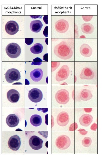
Erythroid cells isolated from slc25a38a+b morphants are morphologically distinct but do not contain any pathological iron deposits compared to cells from control morphants.GFP positive cells were isolated by fluorescence activated cell sorting (FACS) from 48 hpf gata1:EGFP embryos, concentrated onto slides using standard cytospin protocols, and stained with Wright-Giemsa staining for gross morphology and erythroid cell identification (left) and Perls’ Prussian Blue for iron deposits (right). While no pathological iron deposits were detected in the slc25a38a+b morphant cells, these cells are larger with less compact nuclei, indicating a different level of maturation compared with control cells. Images are of randomly selected cells from the same experiment. The experiment was replicated three times with similar results. |
| Fish: | |
|---|---|
| Knockdown Reagents: | |
| Observed In: | |
| Stage: | Long-pec |