
FIGURE
Fig. 7
- ID
- ZDB-FIG-160226-70
- Publication
- Gonsar et al., 2016 - Temporal and spatial requirements for Nodal-induced anterior mesendoderm and mesoderm in anterior neurulation
- Other Figures
- All Figure Page
- Back to All Figure Page
Fig. 7
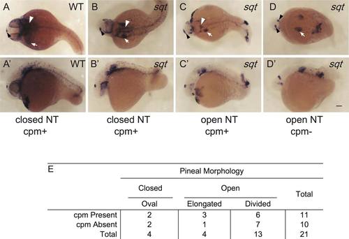
Correlation between cephalic paraxial mesoderm and neural tube closure. (A,A′) In WT embryos, col9a2 is expressed in the cpm (white arrowheads) and the ears (white arrow) and the otx5 expressing pineal anlage (black arrowheads) is oval shaped indicating a closed neural tube. (B–D′) sqt embryos with different combinations of cpm deficiencies and neural tube phenotypes. (A–D) Dorsal views with anterior to the left and (A′–D′) lateral views with anterior to the left and dorsal to the top. Images with the same letter are of the same embryo. (E) Complete set of quantitative data. Scale bar: 100 µm. |
Expression Data
| Genes: | |
|---|---|
| Fish: | |
| Anatomical Terms: | |
| Stage: | Prim-5 |
Expression Detail
Antibody Labeling
Phenotype Data
| Fish: | |
|---|---|
| Observed In: | |
| Stage: | Prim-5 |
Phenotype Detail
Acknowledgments
This image is the copyrighted work of the attributed author or publisher, and
ZFIN has permission only to display this image to its users.
Additional permissions should be obtained from the applicable author or publisher of the image.
Full text @ Genesis