|
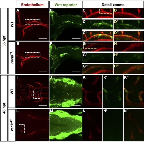
The cerebrovascular expression of the transgenic reporter of canonical Wnt signaling Tg(7xTCF-Xla.Siam:GFP)ia4 is specifically lost in reck y72 mutant embryos. (A-N′′) Confocal images (anterior, left) of 36 and 48hpf Hbs from WT and reck y72. Views: lateral (A-H′′; dorsal, up); dorsal (I-N′′; right side, up). Endothelium, red [Tg(kdrl:RFP)s896]; Wnt reporter, green [Tg(7xTCF-Xla.Siam:GFP)ia4]. In the WT, Wnt reporter expression highlights the PHBCs (A-D′′), CtAs (A-D′′,I-K′′) and additional non-vascular tissues. However, in reck y72 mutants, expression of the Wnt reporter is undetectable in PHBCs (E-H′′,L-N′′) and occasional CtAs (N-N′′), but is present elsewhere. Detail zooms: Regions in white dashed boxes (A,E,I,L) shown in C-C′′,G-G′′,K-K′′ and N-N′′, respectively; regions in yellow dashed boxes (C,G) shown in D-D′′ and H-H′′, respectively. See also Fig. 8.
|