
Fig. 5
- ID
- ZDB-FIG-160212-49
- Publication
- Beerman et al., 2015 - Direct In Vivo Manipulation and Imaging of Calcium Transients in Neutrophils Identify a Critical Role for Leading-Edge Calcium Flux
- Other Figures
- All Figure Page
- Back to All Figure Page
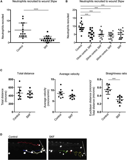
Inhibition of Calcium Channels Disrupts Neutrophil Directionality during Recruitment (A–D) After caudal fin amputation, Tg(LysC:GFP) or Tg(LysC:dsRed) larvae were immediately treated with vehicle alone (control) or 20 µM SKF 96365 (SKF), followed by quantification of neutrophil recruitment 3 hr post-wounding (A and B). (A) Error bars are mean ± SD. Mann-Whitney test p < 0.0001. (B) Groups of larvae were placed in control treatments for 10, 20, or 30 min before replacement of media with SKF for the remainder. Error bars are mean ± SD. One-way ANOVA followed by Tukey’s multiple comparisons test p < 0.001. Each experiment was carried out at least twice. (C and D) Immediately after wounding, larvae were mounted in agarose, immersed with either control or SKF, then imaged with time-lapse microscopy. (C) For the neutrophils tracked, graphs show their total distance traveled, average velocity, and straightness ratio. Total distance and straightness ratio error bars are mean ± SD, and average velocity error bars are mean ± SEM. t test, p = 0.0002. For each group, n = 8 cells from two to three larvae. (D) Final frames from Movie S5 show the tracks of individual neutrophils (quantified in C) near the amputated fin (approximated by the white line). See also Movie S5. |