
FIGURE
Fig. S5
- ID
- ZDB-FIG-160211-23
- Publication
- Abu-Siniyeh et al., 2016 - The aPKC/Par3/Par6 polarity complex and membrane order are functionally inter-dependent in epithelia during vertebrate organogenesis
- Other Figures
- All Figure Page
- Back to All Figure Page
Fig. S5
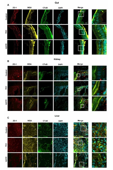
Tissue morphology of gut (A), kidney (B) and liver (C) in sterol 6 dpf zebrafish larvae. Six dpf larvae was left untreated (Control), enriched in 7-ketocholesterol (7KC, 100 µm, 30 min) or depleted of cholesterol with methyl-β-cyclodextrin (mβCD, 2.5 mm, 40 min) as in Figure 3, fixed and stained with antibodies against the tight junction marker ZO-1 (red), WGA (yellow) to mark the cell surface, CTxB (green) to label GM1-rich membranes and DAPI (blue) to visualize cell nuclei. Scale bar in merged images is 20 µm; scale bar in zoomed merged images is 5 µm. |
Expression Data
Expression Detail
Antibody Labeling
Phenotype Data
Phenotype Detail
Acknowledgments
This image is the copyrighted work of the attributed author or publisher, and
ZFIN has permission only to display this image to its users.
Additional permissions should be obtained from the applicable author or publisher of the image.
Full text @ Traffic