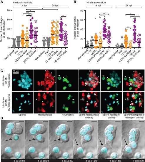
Phagocytes are recruited to the site of infection after spore injection into the hindbrain and interact with spores in vivo. Macrophage (A) and neutrophil recruitment (B) to the site of spore injection in the hindbrain ventricle of Tg(mpeg1:G/U:NfsB-mCherry) and Tg(mpx:GFP) zebrafish larvae, respectively, was observed at 4 and 24h.p.i. Pooled data presented were obtained from three independent experimental repeats with 10 larvae each. (C) Spores can be observed inside macrophages (indicated by asterisks) and neutrophils (indicated by arrowheads) after injection of viable and non-viable spores in the hindbrain ventricle of Tg(mpeg1:G/U:NfsB-mCherry/mpx:GFP) larvae (representative images at 10h 45min and 1h 5min, respectively; z-stack: 15 sections every 7.3µm; scale bar: 20µm). (D) Several subsequent events of spore phagocytosis were also observed in an undefined phagocyte in vivo (arrow indicates next spore to be phagocytosed; time is expressed as h:min:s; scale bar: 4µm; see also Movie 1).
|
