
Fig. 1
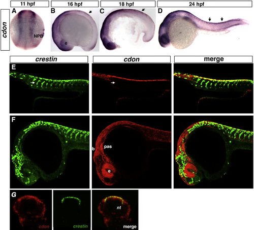
cdon is expressed in the NPB and premigratory trunk NCCs. Dorsal view and anterior to the top (A), and lateral views, anterior to the left (B)–(F). ISH of 11, 16, 18, and 24 hpf stage wildtype embryos. (A)–(D) cdon is expressed in the neural plate border (NPB) and dorsal neural tube (arrows). (E) and (F) Lateral view confocal single optical plane micrograph of double fluorescent ISH of 24 hpf wildtype embryos for crestin (green) and cdon (red). cdon is expressed in premigratory NCCs in the trunk (yellow) but not in the migratory streams (arrows, E) and widely expressed in cranial region including the brain, eye, and pharyngeal arches (F). 14 µm cryosections of double FISH embryos suggests that cdon and crestin colocalize in the dorsal neural tube/neural crest staging area in the trunk (G). b, brain; e, eye; NPB, neural plate border; NT, neural tube; pas, pharyngeal arches. |
Reprinted from Developmental Biology, 407(2), Powell, D.R., Williams, J.S., Hernandez-Lagunas, L., Salcedo, E., O'Brien, J.H., Bruk Artinger, K., Cdon promotes neural crest migration by regulating N-cadherin localization, 289-99, Copyright (2015) with permission from Elsevier. Full text @ Dev. Biol.