
Fig. 6
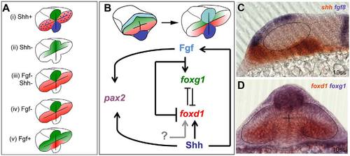
Opposing roles for Fgfs and Shh in the control of optic vesicle patterning. (A) Schematics of foxg1 (green) and foxd1 (red) expression in optic vesicles following manipulations of Fgf and Hh signals. The implications below are based on the ability of Foxg1 to repress foxd1 expression and Foxd1 to repress foxg1 expression. (i) Shh gain of function: loss of foxg1 and gain of foxd1 in nasal retina. This implies that Shh signalling promotes foxd1 expression and/or inhibits foxg1 expression. Blue shading and dots represent Shh expression. (ii) Shh loss of function: loss of foxd1 in temporal retina. This implies that Shh promotes foxd1 expression but is not required for repression of foxg1. (iii) Combined loss of Shh and Fgf: loss of foxg1 and gain of foxd1 in nasal retina. This implies that either unknown signals (grey arrow in B) promote foxd1 expression in absence of Shh or that repressors (such as Fgf itself) are removed in this situation. The result also implies that Fgf is required for the repression of foxd1 in temporal retina (shown in ii), and that this repression is independent of Foxg1 (which is not expressed in temporal retina). (iv) Loss of Fgf expression: loss of foxg1 and gain of foxd1 in nasal retina. This implies that Fgf promotes foxg1 and/or inhibits foxd1 in nasal retina. (v) Gain of Fgf function: gain of foxg1 expression and loss of foxd1 expression in temporal retina (data taken from Picker et al., 2009). This implies that Fgf promotes foxg1 expression and/or inhibits foxd1 expression. (B) Proposed regulatory interactions that could explain the retinal nasotemporal phenotypes shown in A, together with data not shown that both Fgf and Shh promote development of pax2+ optic stalk identity in the proximal optic vesicle. As stated in the main text, the regulatory interactions leading to nasotemporal patterning occur from neural plate stage onwards. (C,D) Images showing the domains of expression in the forebrain of genes encoding the signals studied (C) and their Fox gene targets (D), as evident from double in situ hybridisation assays of 10ss embryos. Dashed lines outline the optic vesicles. |