
Fig. 3
- ID
- ZDB-FIG-151207-3
- Publication
- Karra et al., 2015 - Myocardial NF-κB activation is essential for zebrafish heart regeneration
- Other Figures
- All Figure Page
- Back to All Figure Page
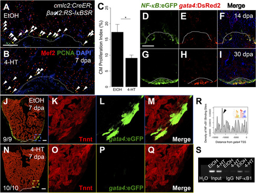
NF-KB modulates cardiomyocyte proliferation and dedifferentiation. (A and B) Section images from cmlc2:CreER; βact2:RS-IκBSR hearts treated with vehicle or 4-HT at 7 dpa. Sections are stained for Mef2 (red) and PCNA (green). Arrowheads indicate Mef2+/PCNA+ nuclei. Dashed line indicates approximate resection plane. (C) Quantification of cardiomyocyte proliferation in cmlc2:CreER; βact2:RS-IκBSR hearts treated with vehicle (n = 15) or 4-HT (n = 17) at 7 dpa. Error bars indicate SEM. *P = 0.004, Mann–Whitney test. (D–I) Sections from NF-KB:eGFP; gata4:DsRed2 hearts at 14 and 30 dpa. (J–Q) Section images from cmlc2:CreER; βact2:RS-IκBSR; gata4:eGFP hearts treated with vehicle (n = 9) or 4-HT (n = 10) at 7 dpa. Inset shows unique high-magnification images for Tnnt, eGFP, and a merge of both channels. (R) Plot for density of putative NFκB1 sites in the gata4 promoter. seqLogo is for the consensus NFκB1 binding site used for analysis. Arrow indicates the region of the promoter with highest density of NFκB1 sites. (S) ChIP-PCR for the region of the gata4 promoter highlighted by arrowhead in R after immunoprecipitation for NFKB1 from cardiac chromatin extracts of cmlc2:CreER; βact2:RS-IκBSR fish treated with vehicle or 4-HT at 7 dpa. (Scale bars: 100 µm.) |