
Fig. S7
- ID
- ZDB-FIG-151102-43
- Publication
- Ghaye et al., 2015 - Progenitor potential of nkx6.1-expressing cells throughout zebrafish life and during beta cell regeneration
- Other Figures
- All Figure Page
- Back to All Figure Page
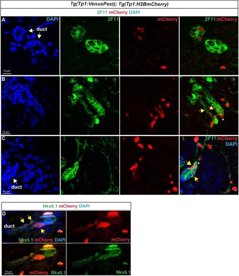
Notch-responsive terminal end duct cells/CACs give rise to other ductal cells. Immunodetection of H2BmCherry and ductal markers in the pancreas of adult Tg(Tp1:VenusPest); Tg(Tp1:H2BmCherry) zebrafish.A–C H2BmCherry+ cells (red) in ductular structures labeled with the ductal (ducts and CACs) marker 2F11 (green). A, B separate channels of Fig. 8b–c. B, C Weak H2BmCherry labeling is present near the extremity of a terminal (or intercalated) duct (yellow arrows). The asterisks identify a CAC at the tip of the duct (strong H2BmCherry labeling). D Comparison of H2BmCherry (red) with endogenous Nkx6.1 (green) showing ductal Nkx6.1+ cells co-expressing H2BmCherry (yellow arrows). |