
Fig. 3
- ID
- ZDB-FIG-151102-3
- Publication
- Blum et al., 2015 - Retinoic acid signaling spatially restricts osteoblasts and controls ray-interray organization during zebrafish fin regeneration
- Other Figures
- All Figure Page
- Back to All Figure Page
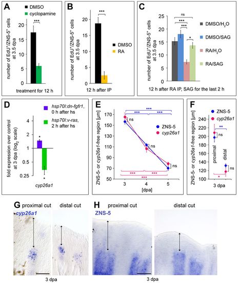
Shha-promoted osteoblast proliferation requires cyp26a1 expression that is restricted to proximal regions by Fgf activity. (A,B) Cyclopamine (A) or prolonged RA (B) treatment downregulates osteoblast proliferation. EdU+/ZNS-5+ cells per section at 3.5dpa. (C) Inhibition of osteoblast proliferation 12h after RA injection can be rescued by concomitant SAG treatment. EdU+/ZNS-5+ cells per section at 3.5dpa. (D) Inhibition of Fgf signaling in hsp70I:dn-fgfr1 fish results in upregulation of cyp26a1 expression. Conversely, activation of Fgf signaling in hsp70I:v-ras fish results in the downregulation of cyp26a1. Transcript levels at 3dpa measured by qPCR. (E-H) ZNS-5- and cyp26a1-free distal domains (double-headed arrows) extend further proximally in regenerates that had been amputated at a more proximal level at 3dpa (F-H), and retracts distally as regeneration proceeds (E). (G) WISH for cyp26a1. (H) IHC for ZNS-5. (E,F) Length of the cyp26a1- or ZNS-5-free distal domain. Data are represented as mean±s.e.m. *P<0.05, **P<0.01, ***P<0.001. ns, not significant; hs, heat shock. Scale bars: 100µm. |