|
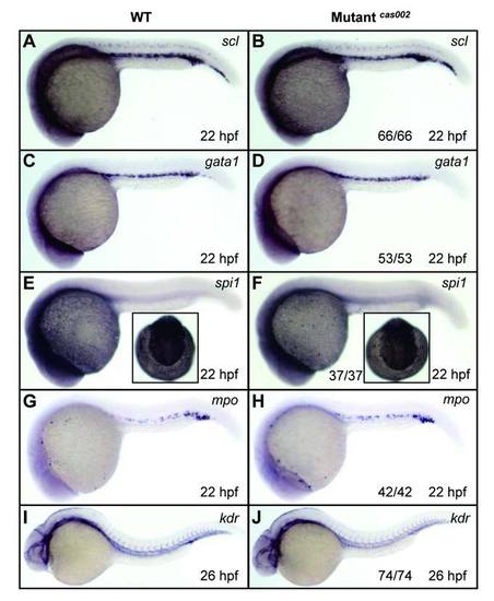
WISH analysis of primitive hematopoiesis and vascular morphogenesis. WT or mutantcas002 embryos were fixed at 22 hpf, followed by WISH analysis of scl, gata1, spi1, mpo expression. Expression of scl (A-B), gata1 (C-D), spi1 (E-F) and mpo (G-H) show no significant difference between wild-type siblings and mutants at 22 hpf. Insets in E-F show dorsal views of the head staining region. Embryos were fixed at 26 dpf, followed by WISH analysis of expression pattern of kdr. Expression of kdr (I-J), which marks vascular endothelial, had no significant difference between siblings and mutants at 26 hpf. The number and percentage of het-het incross embryos with the in situ pattern are listed at the bottom of the Mutantcas002 panels.
|