
Fig. 8
- ID
- ZDB-FIG-151008-1
- Publication
- Aspatwar et al., 2015 - Inactivation of ca10a and ca10b Genes Leads to Abnormal Embryonic Development and Alters Movement Pattern in Zebrafish
- Other Figures
- All Figure Page
- Back to All Figure Page
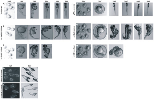
The ca10a and ca10b genes play an important role during embryonic development in zebrafish. Left panel: Developmental images of ca10a and ca10b morphant zebrafish injected with two different sets of antisense MOs (MO1 and MO2) over the period of 0 hpf to 5 dpf. A) The top row images show 0 hpf embryos and lateral views of 1–5 dpf uninjected zebrafish larvae with a normal development of organs,. B) ca10a morphant zebrafish injected with 200 µM translational blocking antisense morpholinos (MO1). C) Images of ca10b morphant zebrafish. D) and E) Images of ca10a and ca10b morphants. The images in first panel of D and E show the 1 dpf larvae with rhodamine fluorescence throughout the larvae (indicating successful MO injection) and second panel (D and E) shows the 5 dpf larvae with abnormal appearance. Right panel: Developmental images of ca10a and ca10b CRISPR mutated fish and gRNA controls. F) The top row images show 0 dpf embryos and lateral views of 1–5 dpf gRNA injected zebrafish larvae (control) with a normal development of organs. G) ca10a mutated zebrafish injected with gRNA and cas9 mRNA targeted to exon 3 (target site shown in Fig 7B). H) ca10b mutated zebrafish injected with gRNA and cas9 mRNA targeted to exon 1(target site shown in Fig 7C). |
| Fish: | |
|---|---|
| Knockdown Reagents: | |
| Observed In: | |
| Stage Range: | Prim-5 to Day 5 |