
FIGURE
Fig. 3
- ID
- ZDB-FIG-150929-12
- Publication
- Lippok et al., 2014 - Pou5f1 protein expression and posttranslational modification during early zebrafish development
- Other Figures
- All Figure Page
- Back to All Figure Page
Fig. 3
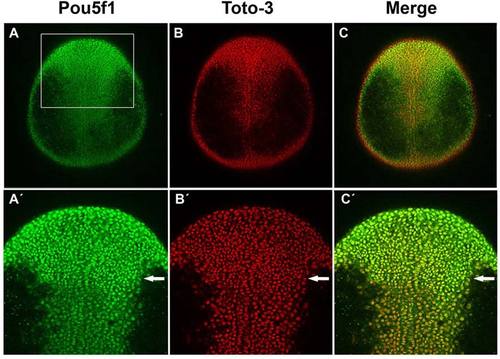
Pou5f1 in dorsal ectoderm cell nuclei in wild-type (WT) embryos at one-somite stage. A–C: Confocal images of anti-Pou5f1 immunofluorescence (green) WT whole-mount embryos at one-somite stage in which all cell nuclei were stained with TOTO-3 (red). A′–C′: Higher magnification of rectangle area in A is shown. Arrows indicate region from which on reduced Pou5f1 levels are detected in the caudal neural plate. Dorsal views, animal pole to the top. |
Expression Data
| Gene: | |
|---|---|
| Fish: | |
| Anatomical Term: | |
| Stage: | 1-4 somites |
Expression Detail
Antibody Labeling
Phenotype Data
Phenotype Detail
Acknowledgments
This image is the copyrighted work of the attributed author or publisher, and
ZFIN has permission only to display this image to its users.
Additional permissions should be obtained from the applicable author or publisher of the image.
Full text @ Dev. Dyn.