
Fig. 5
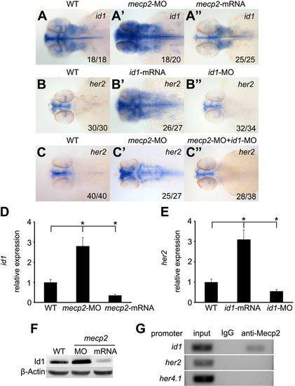
mecp2 depletion directly increases id1 expression that leads to ectopic her2 expression. (A,A′′) RNA in situ hybridization revealed that id1 increased in the developing brain of mecp2 morphants (A′), but decreased in embryos injected with mecp2 mRNA (A′′) compared with wild-type (WT) embryos (A) at 48hpf. (B,B′′) her2 increased in embryos injected with id1 mRNA (B′), but but decreased in id1 morphants (B′′) compared with controls (B) at 48hpf. (C,C′′) her2 increased in mecp2 morphants (C′) and this was rescued to the normal level by co-injecting id1-MO (C′′) compared with wild-type embryos (C). Dorsal views with anterior to the left; numbers in the bottom right corner represent the number of embryos showing the indicated phenotype/total embryos examined. (D,E) Quantitative real-time PCR analysis of id1 expression in wild-type embryos, mecp2 morphants, and embryos injected with mecp2 mRNA, normalized to gapdh (D). her2 expression was also analyzed in wild-type embryos, embryos injected with id1 mRNA and id1 morphants (E). The data represent the mean±s.d. of three independent experiments. Statistics for all groups were calculated by comparison with the wild-type (WT) group. *P<0.05. (F) Western blot revealed that Id1 protein was upregulated in mecp2 morphants, and downregulated in embryos injected with mecp2 mRNA. (G) ChIP assays were performed using an antibody against Mecp2 or IgG control. Semi-quantitative PCR was used to evaluate the promoter occupancy of id1, her2, and her4.1 by Mecp2 protein. Note direct binding of MeCP2 to the id1, but not her2 and her4.1, promoters. One of three independent experiments is shown. |
| Genes: | |
|---|---|
| Fish: | |
| Knockdown Reagents: | |
| Anatomical Terms: | |
| Stage: | Long-pec |