
Fig. 2
- ID
- ZDB-FIG-150805-8
- Publication
- Compagnon et al., 2014 - The Notochord Breaks Bilateral Symmetry by Controlling Cell Shapes in the Zebrafish Laterality Organ
- Other Figures
- All Figure Page
- Back to All Figure Page
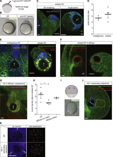
The Notochord Affects KV Cell Shapes (A) Single blastomere injection of constitutively active acvr1b (acvr1b) mRNA at the 32/64-cell stage (1.75/2 hpf) leads to the formation of ectopic KV at the end of gastrulation; mRNA of h2afva fused to a fluorescent protein (FP) is coinjected to label nuclei in the progeny of the injected blastomere. (B) Control (left) and acvr1b-mRNA-injected Tg(sox17:GFP) embryo at 6 ss (12 hpf); white arrow, endogenous KV; purple arrow, ectopic KV. (C) Ectopic KV induced in a Tg(sox17:GFP; actb2:LMA-tdTomato) 6 ss embryo (12 hpf); left panel, 3D rendering; right panel, single plane; arrow points toward the half of the ectopic organ with the highest nuclei density. (D) Maximal nuclei density ratio between organ halves in endogenous (n = 3 embryos) and ectopically (n = 5 embryos) located KV with comparable average cell apical surfaces (between 100 and 120 µm2) in Tg(sox17:GFP) embryo at 6 ss (12 hpf); mean ± SEM. (E) Endogenous (left panel) and ectopic (right panel) KVs in Tg(Ola.Actb:Has.HRAS-EGFP; -1ntla:CFP) embryos at 6 ss (12 hpf); arrow points toward the half of the organ with the highest nuclei density. (F) Ectopic KVs in MZoep; Tg(Ola.Actb:Has.HRAS-EGFP) embryos at 6 ss (12 hpf) located between the yolk and the epiblast (left panel) and between the enveloping layer and the epiblast (right panel). (G) Ectopic KV in a MZoep; Tg(Ola.Actb:Has.HRAS-EGFP) embryo at 6 ss (12 hpf) adjacent to a coinduced notochord (labeled by H2afva-mcherry, yellow dashed line); arrow points toward the half of the ectopic organ with the highest nuclei density. (H) Maximal nuclei density ratio between organ halves in ectopic KV at 6 ss (12 hpf) in control Tg(sox17:GFP) (n = 5 embryos), MZoep; Tg(Ola.Actb:Has.HRAS-EGFP) (n = 4 embryos), and MZoep; Tg(Ola.Actb:Has.HRAS-EGFP) with coinduced notochord (n = 2 embryos) embryos with comparable average cell apical surfaces (between 100 and 120 µm2); mean ± SEM; p < 0.05 (Mann-Whitney test). (I) Single marginal blastomere injection of acvr1b mRNA at the 16-cell stage (2 hpf) leads to the formation a secondary axis, whose notochord contacts the posterior side of endogenous KV at the end of gastrulation; upper panel schematizes a gastrulating embryo with both endogenous and induced axes; lower left panel shows a Tg(sox17:GFP) embryo at 80% epiboly (8.5 hpf) injected with Acvr1b mRNA at the 16-cell stage (2 hpf) with both primary (purple arrow) and secondary (yellow arrow) axes. (J) KV adjacent to the notochord from both the primary (purple dashed line) and secondary (yellow dashed line) axes in a Tg(sox17:GFP; actb2:LMA-tdTomato) embryo at 6 ss (12 hpf). (K) α-Acetylated tubulin staining of KV in Tg(sox17:GFP) control embryos (upper panels) at 6 ss (12 hpf) and in similar staged embryos with a secondary axis where KV is adjacent to the notochord from both the primary (white dashed line) and secondary (yellow dashed line) axes; panels at the right show the corresponding cilia distribution based on z-projections. An, animal pole; Veg, Vegetal pole; noto, notochord; Ac. Tubulin, acetylated tubulin; ctrl, control. Scale bars for (C), (E), (F), (G), (J), and (K), 20 µm. See also Figure S2B. |
Reprinted from Developmental Cell, 31, Compagnon, J., Barone, V., Rajshekar, S., Kottmeier, R., Pranjic-Ferscha, K., Behrndt, M., Heisenberg, C., The Notochord Breaks Bilateral Symmetry by Controlling Cell Shapes in the Zebrafish Laterality Organ, 774-783, Copyright (2014) with permission from Elsevier. Full text @ Dev. Cell