
Fig. 2
- ID
- ZDB-FIG-150716-9
- Publication
- Rau et al., 2015 - Abnormal splicing switch of DMD's penultimate exon compromises muscle fibre maintenance in myotonic dystrophy
- Other Figures
- All Figure Page
- Back to All Figure Page
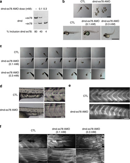
Exclusion of dmd exon 78 in zebrafish impairs skeletal muscle development. (a) RT–PCR of dmd exon 78 performed on total RNA extracts isolated from whole control and dmd Δ78 embryos (48 hpf). (b) Dose-dependant phenotype of dmd Δ78 embryos: control embryos (CTL) compared with moderate and severe affected dmd Δ78 morphants at 48 hpf (scale bar, 1 mm). (c) Touch-evoked escape test of control embryos compared with moderate and severe dmd Δ78 morphants (1 image/0.2 s; scale bar, 1 mm). (d) Abnormal myoseptum U-shape in dmd Δ78 morphants compared with V-shape in control embryos at 48 hpf (scale bar, 250 and 100 µm). (e) Dystrophin immunostaining (MANDRA1 antibody) of control embryos compared with dmd Δ78 morphants at 48 hpf. (f) Slow myosin immunostaining of control embryos compared with moderate and severe affected dmd Δ78 morphants at 48 hpf (scale bar, 50 and 10 µm). |
| Gene: | |
|---|---|
| Antibody: | |
| Fish: | |
| Knockdown Reagent: | |
| Anatomical Term: | |
| Stage: | Long-pec |
| Fish: | |
|---|---|
| Knockdown Reagent: | |
| Observed In: | |
| Stage: | Long-pec |