
Fig. 1
- ID
- ZDB-FIG-150716-14
- Publication
- Xiao et al., 2015 - High-resolution live imaging reveals axon-glia interactions during peripheral nerve injury and repair in zebrafish
- Other Figures
- All Figure Page
- Back to All Figure Page
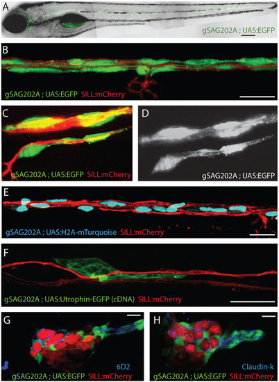
Tg[gSAGFF202A] is a specific Gal4 driver in Schwann cells. (A) EGFP expression pattern at 5dpf by Tg[gSAGFF202A;UAS:EGFP]. (B-D) Triple transgenic Tg[gSAGFF202A;UAS:EGFP;SILL:mCherry] at 5dpf show that EGFP(+) cells form tubes wrapping around an mCherry(+) axon by confocal (B) and lattice light-sheet (C,D) imaging. (E) A triple-transgenic larva (Tg[gSAGFF202A;UAS:H2A- mTurquoise;SILL:mCherry]) at 5dpf. Nuclei of cells that are in intimate contact with lateralis afferent axons are shown in blue along the trunk. (F) Marking of individual cell shows UtrCH-EGFP expression in a 4-dpf Tg[gSAGFF202A;SILL:mCherry] double-transgenic fish injected with UAS:UtrCH-EGFP construct. (G) The marker 6D2 labels EGFP-expressing cells in a lateral plane of posterior ganglion. (H) The marker Claudin-k labels EGFP-expressing cells in a lateral plane of posterior ganglion. In all figures, dorsal is up and anterior is left. Scale bars: 150µm (A) and 10µm (B-H). |
| Genes: | |
|---|---|
| Antibody: | |
| Fish: | |
| Anatomical Terms: | |
| Stage: | Day 5 |