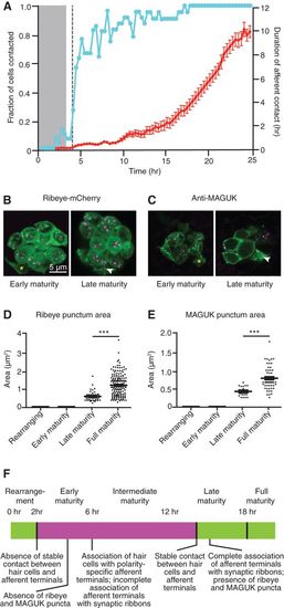
Hair cells form synapses 2–15 h after mitosis. (A) The fraction of nascent hair cells making contact with afferent terminals (blue) rises briskly following cellular rearrangement. Although the contacts are transitory at the outset, their duration increases progressively until, by 15 h after mitosis, the slope of the line approaches unity, indicating complete stability (red). Time is denoted in hours after mitosis. The gray band marks the period of cellular rearrangement, and the vertical dashed line marks the emergence of projections. N = 30. (B–E) Ribeye-mCherry fusion protein (B,D) and membrane-associated guanylate kinase (MAGUK) (C,E) puncta (arrowheads) appear in late maturity and full maturity hair cells (magenta) but are absent from rearranging cells and early maturity cells (asterisks in B,C). For D and E, P < 0.0001; for D, N = 8, 34, 47, and 176 measurements for the respective hair cell stages; for E, N = 8, 16, 16, and 58 measurements. (F) A time line of hair cell differentiation. The magenta box denotes the extension of projections from nascent hair cells from shortly after their rearrangement until stable synapses have formed (see Figs. 2-4).
|
