
FIGURE
Fig. S3
- ID
- ZDB-FIG-150528-17
- Publication
- Lenard et al., 2015 - Endothelial Cell Self-fusion during Vascular Pruning
- Other Figures
- All Figure Page
- Back to All Figure Page
Fig. S3
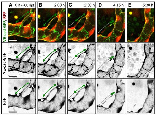
Final steps of pruning type I. A small, lumenized branch is made of two cells (A). Lumen collapses when the branch is still multicellular (B); after lumen collapse, cells move away from each other, and the cell–cell contact surface shrinks (C). The last cell–cell contact (D, green arrow) is eventually resolved completely as the last cytoplasmic extention of the bridging cell detaches from the major branch (E). See also S9 Movie. |
Expression Data
Expression Detail
Antibody Labeling
Phenotype Data
Phenotype Detail
Acknowledgments
This image is the copyrighted work of the attributed author or publisher, and
ZFIN has permission only to display this image to its users.
Additional permissions should be obtained from the applicable author or publisher of the image.
Full text @ PLoS Biol.