
Fig. 6
- ID
- ZDB-FIG-150526-6
- Publication
- Ye et al., 2015 - glucagon is essential for alpha cell transdifferentiation and beta cell neogenesis
- Other Figures
- All Figure Page
- Back to All Figure Page
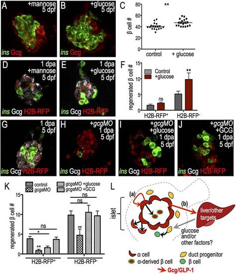
Differential regulation of β cell progenitor pools by glucagon and glucose. (A,B) Confocal projections of Tg(ins:CFP-NTR) islets with 1-day treatment with mannose (osmolality control, A) or glucose (B) and staining for CFP (insulin, green) and glucagon (red). (C) Quantification of insulin+ cells showed that glucose treatment increases β cell mass (n=18). (D,E) Confocal projections of 5-dpf/1-dpa H2B-RFP mRNA-injected islets that were treated with mannose (D) or glucose (E) during regeneration and stained for CFP (green) and glucagon (white). (F) Quantification of H2B-RFP+ and H2B-RFP β cells in control (n=7) and glucose-treated (n=7) islets shows a specific increase in the H2B-RFP population. (G-J) Confocal projections of 5-dpf/1-dpa Tg(ins:CFP-NTR) islets injected with H2B-RFP mRNA alone (G) or co-injected with gcgaMO (H-J). Samples were treated with vehicle (G,H), glucose (I) or glucagon (J). (K) Quantification of H2B-RFP+ and H2B-RFP cells from islets in control (n=27), gcgaMO (n=15), gcgaMO+glucose (n=17), gcgaMO+glucagon (n=22) treatments. Only glucagon rescued H2B-RFP+ β cell regeneration. (L) Model integrating distinct roles of glucagon in β cell formation. Loss of β cells triggers α cell proliferation and activation of glucagon. Increased output of glucagon and GLP-1 drives regeneration from two sources: (a) glucagon/GLP-1 acts autonomously in α cells to permit their transdifferentiation to β cells; and (b) glucagon/GLP-1 acts non-autonomously through intermediates (e.g. liver-derived glucose) to drive β cell formation from duct-associated progenitors. *P≤0.05, **P≤0.01; ns, not significant (Student′s t-test in C,F; one-way ANOVA followed by Tukey′s post-hoc test in K). |
| Gene: | |
|---|---|
| Antibody: | |
| Fish: | |
| Condition: | |
| Anatomical Terms: | |
| Stage: | Day 5 |