|
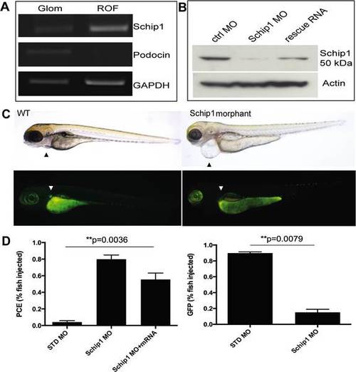
Schip1 is expressed in zebrafish pronephros and its inactivation leads to pericardial edema and loss of podocyte specific GFP-expression (A) To confirm the presence of Schip1 in zebrafish, pronephroi were microdissected from the zebrafish line expressing the GFP under podocin promoter. RT-PCR from pronephros (Glom) and rest-of-fish (ROF) fractions shows the signal for Schip1 in pronephros. Podocin was used as the positive control to validate the purity of the pronephros fraction and GAPDH as the loading control. (B) Morpholino injection in zebrafish resulted in the reduction of Schip1 protein as confirmed by Western blot. An increase in Schip1 protein is detected in fish rescued by coinjection with mouse full-length Schip1 mRNA. Zebrafish Schip1 protein band 50kDa. Actin-beta used as loading control. (C) Inactivation of Schip1 led to development of pericardial edema in 96 hpf morphant embryos (upper panels, arrowheads). In podocin-GFP zebrafish line, Schip1 injection caused loss of GFP signal in pronephros (lower panels, arrowheads). (D) Graphs showing quantification of the zebrafish phenotypes. Data presented as mean with SEM of several experiments. WT-wild type, MO-morpholino, PCE-pericardial edema, ctrl-control, GFP-green fluorescent protein.
|