
FIGURE
Fig. 5
- ID
- ZDB-FIG-150506-29
- Publication
- Dirian et al., 2014 - Spatial Regionalization and Heterochrony in the Formation of Adult Pallial Neural Stem Cells
- Other Figures
- All Figure Page
- Back to All Figure Page
Fig. 5
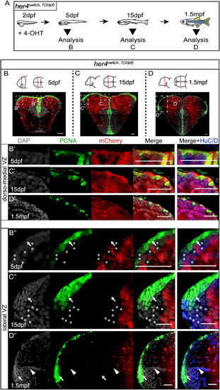
Pallium Development Occurs in Two Heterochronic Waves (A) Experimental design. (B–D) Cross-sections of the telencephalon in her4switch,T(2 dpf) animals at 5 dpf (B), 15 dpf (C), and 1.5 mpf (D) stained as indicated. Arrows and asterisks highlight, respectively, the PCNA+/mCherry- progenitors and the first neurons of the lateral pallium. Arrowheads indicate the lateral pallial sulcus. See also Figure S5. |
Expression Data
Expression Detail
Antibody Labeling
Phenotype Data
Phenotype Detail
Acknowledgments
This image is the copyrighted work of the attributed author or publisher, and
ZFIN has permission only to display this image to its users.
Additional permissions should be obtained from the applicable author or publisher of the image.
Reprinted from Developmental Cell, 30(2), Dirian, L., Galant, S., Coolen, M., Chen, W., Bedu, S., Houart, C., Bally-Cuif, L., Foucher, I., Spatial Regionalization and Heterochrony in the Formation of Adult Pallial Neural Stem Cells, 123-36, Copyright (2014) with permission from Elsevier. Full text @ Dev. Cell