
FIGURE
Fig. 6, S3
- ID
- ZDB-FIG-150420-33
- Publication
- Heermann et al., 2015 - Eye morphogenesis driven by epithelial flow into the optic cup facilitated by modulation of bone morphogenetic protein
- Other Figures
- All Figure Page
- Back to All Figure Page
Fig. 6, S3
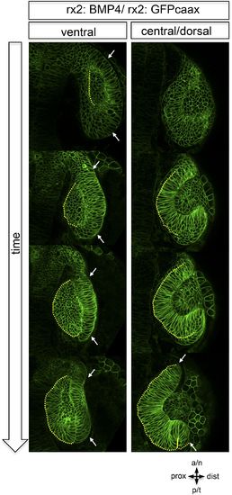
Dorsal view on optic cup development of an rx2::BMP4 embryo over time at ventral vs central/ dorsal levels. The yellow dotted line indicates the border between the lens-facing and the lens-averted domain. Remarkably, the lens-averted domain is not integrated into the optic cup (compare to Figures 1 and 2). Notably, an altered morphology of the ventral optic vesicle can be observed showing the domain which is not going to be integrated (arrows). These data are derived from imaging data (start at 19 hpf) which is presented in Video 7. |
Expression Data
Expression Detail
Antibody Labeling
Phenotype Data
Phenotype Detail
Acknowledgments
This image is the copyrighted work of the attributed author or publisher, and
ZFIN has permission only to display this image to its users.
Additional permissions should be obtained from the applicable author or publisher of the image.
Full text @ Elife