
Fig. 1
- ID
- ZDB-FIG-150420-2
- Publication
- Bassi et al., 2015 - Optical tomography complements light sheet microscopy for in toto imaging of zebrafish development
- Other Figures
- All Figure Page
- Back to All Figure Page
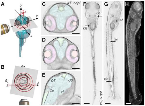
Optical tomography principles and results. (A) Scheme of the acquisition system. During the measurement the specimen is translated and rotated through the focal plane of the detection objective lens (x,y). The specimen is sampled along a spiral. (B) Scheme of the system from the top. The spiral is formed on the transverse section of the specimen. The detection objective′s depth of field, δz, is highlighted in red. (C-E) Transverse (C), coronal (D) and sagittal (E) slices of a wild-type 2dpf zebrafish head obtained in vivo with optical tomography (reconstructed virtual sections). Segmented head organs: retina (pink), eye lens (orange), brain ventricles (green), brain (cyan). Annotated brain domains: optic tectum (OT), hypothalamus (H), cerebellum (Ce) and olfactory bulb (OB). (F,G) Coronal (F) and sagittal (G) slices of a 5dpf zebrafish. SB, swim bladder; OC, otic capsule; Li, liver; So, somites; No, notochord. (H) Lateral view of the 3D reconstructed sample. Scale bars: 100µm. |