
Fig. 6
- ID
- ZDB-FIG-150406-15
- Publication
- Tzung et al., 2015 - Early Depletion of Primordial Germ Cells in Zebrafish Promotes Testis Formation
- Other Figures
- All Figure Page
- Back to All Figure Page
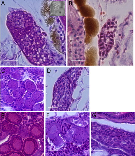
Hematoxylin and Eosin Staining of WT and PGC-Depleted Zebrafish Gonads at Different Developmental Stages (A) WT gonads at 15 dpf contained meiotic germ cells indicating oogonia (white box, inset). (B) PGC-depleted gonads at 15 dpf did not contain differentiated germ cells. (C and D) At 23 dpf, WT (C) and PGC-depleted gonads (D) are shown. Perinucleolar oocytes were only identified in the WT gonad (arrow). PGC-depleted gonads contained germ cells with one to several nucleoli. (E) WT gonads at 28 dpf showed slightly packed oocytes at the perinucleolar stage (arrow). (F and G) At 28 dpf, some PGC-depleted gonads have developed ovarian structures with perinucleolar oocytes (F, arrow), whereas others were similar histologically to PGC-depleted gonads of 23 dpf (compare G and D). Scale bars represent 20 µm. See also Figures S3 and S4. |