
Fig. 3
- ID
- ZDB-FIG-150402-3
- Publication
- Kok et al., 2015 - Reverse Genetic Screening Reveals Poor Correlation between Morpholino-Induced and Mutant Phenotypes in Zebrafish
- Other Figures
- All Figure Page
- Back to All Figure Page
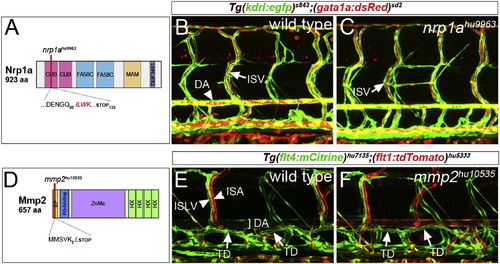
Normal Vascular and Lymphatic Development in nrp1a and mmp2 Mutant Embryos (A) Schematic of Nrp1a domain structure, indicating position of nrp1ahu9963. CUB, CUB domain; FA58C, coagulation factor 5/8 C-terminal domain; MAM, MAM domain; and DUF3481, domain of unknown function. (B and C) Confocal micrograph of embryos bearing Tg(kdrl:egfp)s843 (green) and Tg(gata1a:dsRed)sd2 (red) transgenes at 5 days postfertilization. (B) Wild-type: DA and ISV are indicated. (C) nrp1ahu9963 mutant: ISV carrying red blood cells is indicated. (D) Schematic of the Mmp2 and mmp2hu10535 allele. SP, signal peptide; PG-binding, peptidoglycan-binding domain; ZnMc, zinc-dependent metalloprotease; and HX, hemopexin-like repeats. (E and F) Confocal micrographs of embryos bearing Tg(flt4:mCitrine)hu7135 (green) and Tg(flt1:tdTomato)hu5333 (red) transgenes. (E) Wild-type: ISLV, intersegmental artery (ISA), DA, and TD are indicated. (F) mmp2hu10535 mutant embryo. |
| Fish: | |
|---|---|
| Observed In: | |
| Stage: | Day 5 |
Reprinted from Developmental Cell, 32(1), Kok, F.O., Shin, M., Ni, C., Gupta, A., Grosse, A.S., van Impel, A., Kirchmaier, B.C., Peterson-Maduro, J., Kourkoulis, G., Male, I., DeSantis, D.F., Sheppard-Tindell, S., Ebarasi, L., Betsholtz, C., Schulte-Merker, S., Wolfe, S.A., Lawson, N.D., Reverse Genetic Screening Reveals Poor Correlation between Morpholino-Induced and Mutant Phenotypes in Zebrafish, 97-108, Copyright (2015) with permission from Elsevier. Full text @ Dev. Cell