
Fig. 7
- ID
- ZDB-FIG-150324-25
- Publication
- Marchesi et al., 2014 - DEPDC1B Coordinates De-adhesion Events and Cell-Cycle Progression at Mitosis
- Other Figures
- All Figure Page
- Back to All Figure Page
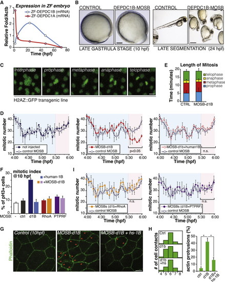
DEPDC1B Controls Mitotic Events in Zebrafish Embryo (A) Zebrafish (ZF) embryos were collected at different times from fertilization (hpf) and levels of depdc1b or depdc1a mRNA expression measured by RT-qPCR. Actin B (Actb) was used as a normalizer. (B) ZF embryos were injected with control or depdc1b splice-blocking morpholino (MOSB) at the one-cell stage. Pictures show representative images of the resulting morphological defects observed at the late gastrula (10 hpf, left) or late segmentation (24 hpf, right) stages. Scale bars represent 125 µm (left) and 500 µm (right). (C–I) Zebrafish embryos were treated as in (B), and mitotic figures were monitored in the embryonic dorsal region of the Tg(h2afva:GFP)kca6 zebrafish transgenic line using confocal microscopy. Representative images are shown in (C).The scale bar represents 10 µm. (D) The graphs show the number of mitotic events (mean ± SD of two experiments) occurring from 4 to 6 hpf. DEPDC1B human mRNA was microinjected together with depdc1b MOSB to rescue embryo defects. (E) Duration of mitotic phases (mean ± SEM) was measured by confocal time-lapse microscopy, after MBT. (F) The graphs show the number of mitotic cells at 10 hpf determined by immunofluorescence upon various treatments (mean ± SEM of two experiments). (G) Cortical actin cytoskeleton was analyzed in the anterodorsal region of embryos treated as in (D), at 10 hpf by FITC-phalloidin staining. Representative images are shown. Red arrows mark thickening and actin protrusions on cell edges. hs-1B, human DEPDC1B mRNA. The scale bar represents 10 µm. (H) Bar graphs show the number of cell contacts (left panel) and actin protrusions observed in two independent experiments (mean ± SEM). Asterisk marks significant values (p < 0.05). (I) Embryos were treated as in (D), with different splice-blocking morpholinos (MOSBs for depdc1b, rhoab, or ptprf) at the one-cell stage. The graphs show the number of mitotic events (mean ± SD of two experiments) occurring from 4 to 6 hpf. |
| Fish: | |
|---|---|
| Knockdown Reagent: | |
| Observed In: | |
| Stage Range: | Bud to Prim-5 |
Reprinted from Developmental Cell, 31, Marchesi, S., Montani, F., Deflorian, G., D'Antuono, R., Cuomo, A., Bologna, S., Mazzoccoli, C., Bonaldi, T., Di Fiore, P.P., Nicassio, F., DEPDC1B Coordinates De-adhesion Events and Cell-Cycle Progression at Mitosis, 420-433, Copyright (2014) with permission from Elsevier. Full text @ Dev. Cell